
AI and Intelligence
Medical Image Analysis
AI-assisted analysis that helps professionals review medical images — highlighting, quantifying and organising, while reading remains a professional act.
Overview
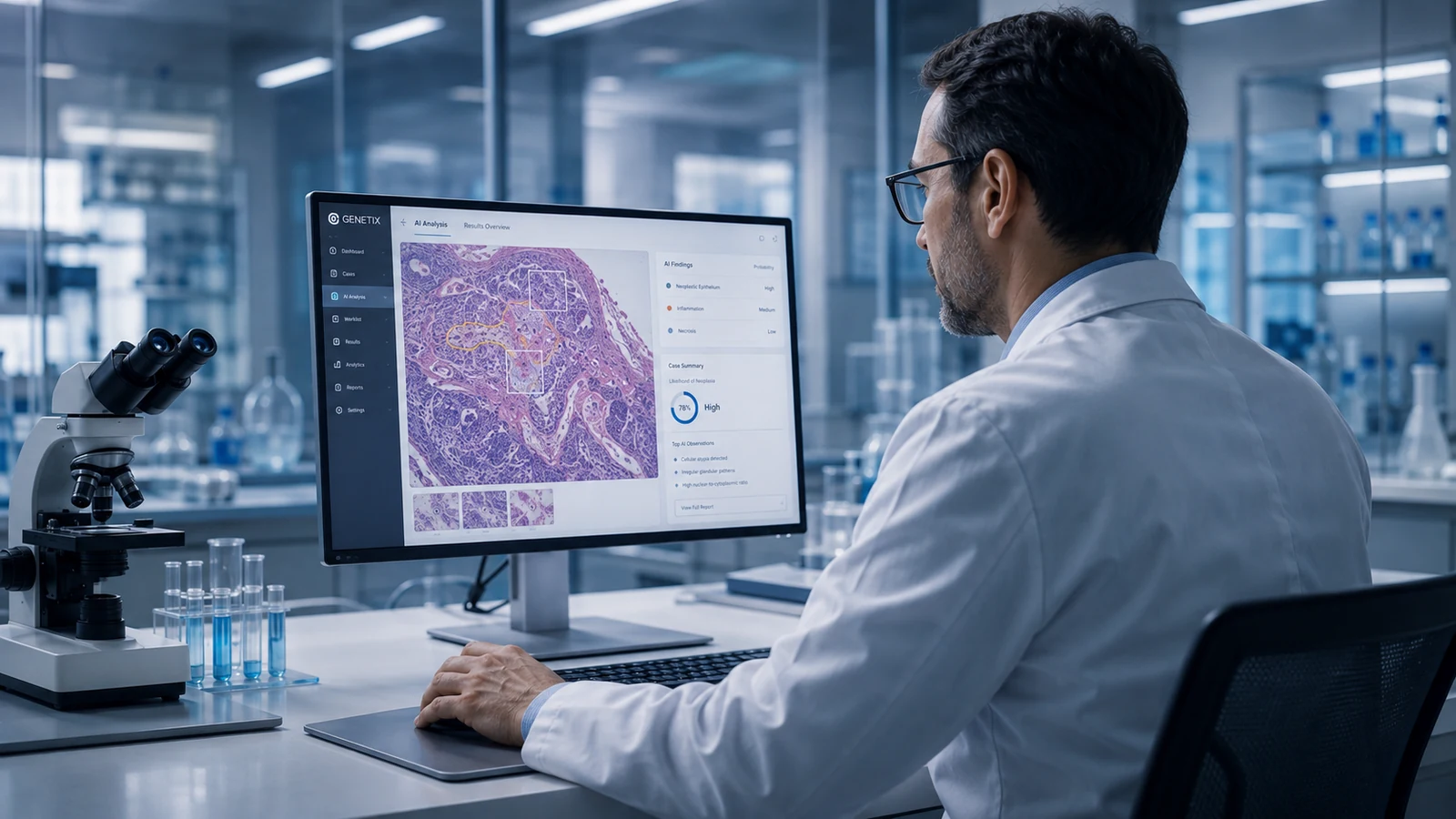
Genetix medical image analysis applies AI models to imaging studies to support professional review: detecting and marking candidate regions for attention, producing consistent quantifications, and helping order the reading worklist. Findings are presented as overlays and structured results alongside the study — clearly marked as AI-assisted analysis.
The professional's read is the outcome; analysis results are inputs to it. Results flow into the record in structured form, so downstream decisions and multidisciplinary discussion can build on them, and how the analysis behaved remains traceable.
Healthcare context
Imaging volumes grow faster than reading capacity. Specialists need support that saves attention rather than competing for it: consistent measurements, prioritisation of worklists, and indications of regions that deserve a closer look.
Intended users
- Radiologists and imaging specialists
- Multidisciplinary teams using imaging results
- Imaging IT and clinical physics
Availability and deployment options are confirmed as part of an initial consultation with our team.
Capabilities
What it provides
Region marking for review
Model-detected candidate regions presented as reviewable overlays on the study.
Consistent quantification
Automated measurements that support comparison over time and between studies.
Worklist support
Configurable ordering of reading worklists based on analysis outcomes.
Structured results to the record
Analysis outputs stored in structured form, linked to the study and the professional read.
Traceable model behaviour
Versioned models with logged behaviour, supporting evaluation and governance.
Example workflow
How it works in practice
- 1
A study arrives
The study is routed to the configured analysis pipeline on receipt.
- 2
Analysis runs and is attached
Overlays and quantifications are attached to the study, marked as AI-assisted.
- 3
The specialist reads
The professional reviews the study with the analysis available — confirming, correcting or discarding its findings.
- 4
The read is reported
The professional report is the clinical result; analysis context stays linked and traceable.
Example workflow for illustration. Actual configurations are defined with each organisation and always keep qualified professionals in control of clinical decisions.
Integration
Part of a connected environment
Image analysis integrates with imaging infrastructure and the record through the integration platform, using structured healthcare data exchange configured per deployment.
Image analysis feeds the intelligence pillar with some of its most concrete outputs and depends on the integration layer to place those outputs where professionals work.
Connects with
- Imaging systems and archives (per deployment)
- Electronic health records
- Clinical decision support
- Audit and model-governance services
Implementation
Introduced with care
Deployments are scoped per use case: modality, pathway, model configuration and evaluation approach are defined with the imaging department. Regulatory and validation requirements are identified per use case and jurisdiction before clinical use.
Responsible use
- Analysis supports the read; it does not produce the diagnosis.
- AI-assisted content is always identifiable as such.
- Use cases requiring formal validation or registration are identified per deployment and jurisdiction; no clinical-performance claims are made outside validated contexts.
Related
Explore further
Get in touch
Talk to our team about Image Analysis.
We can walk through the capability in the context of your organisation, your systems and your priorities.